
Ossification of the Posterior Longitudinal Ligament (OPLL)
Blog
August 3, 2019Know About Cervical Artificial Disc Replacement Surgery
August 26, 2019
Ossification of the posterior longitudinal ligament (OPLL) is a condition in which a flexible
the structure known as the posterior longitudinal ligament becomes thicker and less flexible.
The posterior longitudinal ligament connects and stabilizes the bones of the spinal column.
It runs almost the entire length of the spine, from the 2nd vertebra in the cervical spine
(neck) all the way down to the sacrum (end of the spine). The ligament is adjacent to the
spinal cord.
In Normal circumstances when posterior longitudinal ligament occupies spinal canal, it does
cause any harm to the spinal cord, but when it gets ossified it hardens like bone and encroach in
space in the spinal canal which normally otherwise occupied by the spinal cord. Thus it causes
compression of the spinal cord giving rise to the condition known as compressive myelopathy
(Affection of spinal cord). OPLL most often occurs at the cervical spine (spine in the neck),
causing Cervical myelopathy. Less often it can affect dorsal spine causing dorsal myelopathy.
Here at The New Bombay Spine Clinic, we specialize in treating OPLL causing a reversal of
myelopathy condition.
Symptoms
OPLL typically begins with no or mild symptoms. Mild symptoms may include mild pain
tingling, and/or numbness in the hands. OPLL can also cause dysesthesia, an unpleasant
sensation that accompanies touch. Sometimes an unpleasant sensation may be present
without any touch. As OPLL progresses, symptoms typically become more severe. If the
ligament takes up valuable space within the spinal canal as it thickens, it may compress
(squeeze) the spinal cord, producing myelopathy. Symptoms of myelopathy (spinal cord
compression) include difficulty walking and difficulty with bowel and bladder control. OPLL
may also cause radiculopathy or compression of a nerve root. Symptoms of cervical
radiculopathy include pain, tingling, or numbness in the neck, shoulder, arm, or hand. The
majority of cases will include a slow progression of symptoms, but in some cases, symptoms
may suddenly become worse after a mild injury. The majority of cases will include a slow
progression of symptoms, but in some cases, symptoms may suddenly become worse after
a mild injury.
Causes and Risk Factors
The causes of OPLL are not fully understood. Genetic, hormonal, environmental, and lifestyle
factors seem to play a role. OPLL is usually detected in men in their 50’s and 60’s.
Tests and Diagnosis
It is most common in individuals with Asian, especially Japanese, ancestry. If a patient
presents with symptoms associated with OPLL, the doctor may order the following
diagnostic procedures:
X-ray (also known as plain films) –a test that uses invisible electromagnetic energy
beams (X-rays) to produce images of bones. Soft-tissue structures such as the spinal
cord, spinal nerves, the disc, and ligaments are usually not seen on X-rays, nor on
most tumors, vascular malformations, or cysts. X-rays provide an overall assessment
of the bone anatomy as well as the curvature and alignment of the vertebral column.
Spinal dislocation or slippage (also known as spondylolisthesis), kyphosis, scoliosis, as
well as local and overall spine balance can be assessed with X-rays. Specific bony
abnormalities such as bone spurs, disc space narrowing, vertebral body fracture,
collapse or erosion can also be identified on plain film X-rays. Dynamic or
flexion/extension X-rays (X-rays that show the spine in motion) may be obtained to
see if there is any abnormal or excessive movement or instability in the spine at the
affected levels.
Computed tomography (CT) scan— a diagnostic imaging procedure that uses a
combination of X-rays and computer technology to produce detailed images of any
part of the body, including the bones, muscles, fat, and organs. CT scans are more
detailed than general X-rays.
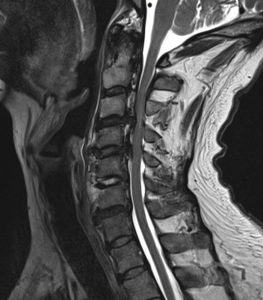
Magnetic resonance (MR) imaging — a diagnostic procedure that uses a combination
of large magnets, radio waves, and a computer to produce detailed images of organs
and structures within the body. MR imaging scans use no radiation. They may not be
possible in patients with certain implants or devices, such as pacemakers or old
aneurysm clips.
Treatments
When symptoms are mild and not progressive, OPLL can be addressed with nonoperative
measures. Nonoperative treatments may include pain medications, anti-inflammatory
medications, anticonvulsants, non-steroidal anti-inflammatory drugs (NSAIDs) and topical
opioids.
However, surgery may be considered if a patient develops signs or symptoms of
myelopathy, such as abnormal reflexes or difficulty walking, or if it is radiographic
evidence of injury or ongoing compression of the spinal cord.
The surgeon may perform any of the following procedures:
Anterior cervical discectomy with fusion (ACDF)
Anterior cervical corpectomy with fusion
Laminectomy
Laminectomy and fusion
Laminoplasty
Combined anterior and posterior approach
The surgeon will determine the best treatment for each patient and situation. Treatment
decisions will depend on a variety of factors, such as the degree of myelopathy, spinal
deformity, and the number of segments involved.